
In 1924, a flickering line on a strip of paper in a Jena hospital room started the field of EEG. A hundred years later, we’re telling that story and revealing why we just named our new update after the city where it all began.
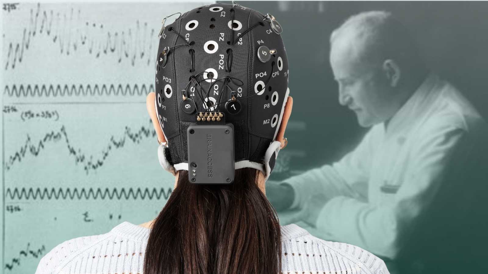
On July 6, 1924, in a room at the University Hospital in Jena, a German psychiatrist named Hans Berger placed electrodes on the scalp of a 17-year-old patient and watched a galvanometer needle twitch.
It wasn’t much to look at. A faint, irregular tremor on a strip of paper. But it was the first time anyone had recorded the electrical activity of a living human brain from outside the skull. Berger didn’t announce it. He didn’t even fully trust it. He spent the next five years quietly repeating the experiment, refining his equipment, and trying to convince himself the signal was real before he dared publish anything.
Box 1: Before Berger
Berger wasn’t working in a vacuum. Decades earlier, Luigi Galvani had shown that electricity could make a frog’s muscle twitch; this was the first hint that biology and electricity were connected. In 1875, the British physiologist Richard Caton went further, recording electrical activity directly from the exposed brains of animals. It was clear the brain produced electrical signals. What nobody had done was catch that signal on a living human, through the scalp, without opening anything up.
That was the problem Berger set out to solve as a psychiatrist. He’d spent years searching for a physical basis for telepathy and psychic phenomena, and kept coming up empty. Frustrated, he turned his instruments toward something more measurable: the brain’s own electrical activity. It was a strange detour that ended up rewriting neuroscience.
The decade nobody believed him
When Berger finally published his findings in 1929, the scientific community mostly shrugged. Some doubted his equipment could detect anything meaningful through the scalp and skull. Others assumed he was picking up artifacts (i.e., muscle twitches, eye movements). Berger kept working anyway, cataloguing the different rhythms he saw: fast, small waves he called beta, and a slower, more prominent rhythm, around 8 to 12 cycles per second, that he named alpha.
The turning point came in 1934, a full decade after that first recording, when the British physiologists Edgar Adrian and B.H.C. Matthews independently confirmed Berger’s results in front of a skeptical scientific audience.
Alpha waves were real. Berger had been right all along, and the rhythm he’d first described is still sometimes called the Berger rhythm today. He’d also given the field its name, coining the term “electroencephalogram” (electric-brain-writing) which is where the “EEG” on every device, paper, and press release since has come from.

From curiosity to clinic and human-computer interaction
Once EEG was validated, it moved fast. Through the 1930s and 40s, researchers realized the technique could do more than confirm a theory: it could diagnose.
- Distinctive spike patterns showed up during epileptic seizures.
- Different rhythms marked the stages of sleep.
- Anesthesiologists found they could track how deeply unconscious a patient was.
Dedicated EEG hardware started appearing in hospitals, and by mid-century, EEG had gone from one psychiatrist’s obsession to a standard tool of clinical neurology.
The next big shift came when researchers stopped asking only what does this signal tell us? and started asking what could this signal let us do? By the 1970s, scientists were exploring whether brain signals could be used to control something external (a cursor, a switch, eventually entire interfaces) and the field of brain-computer interfaces (BCI) was born.
EEG stopped being purely diagnostic and became interactive.
A hundred years later: from Jena to Jena
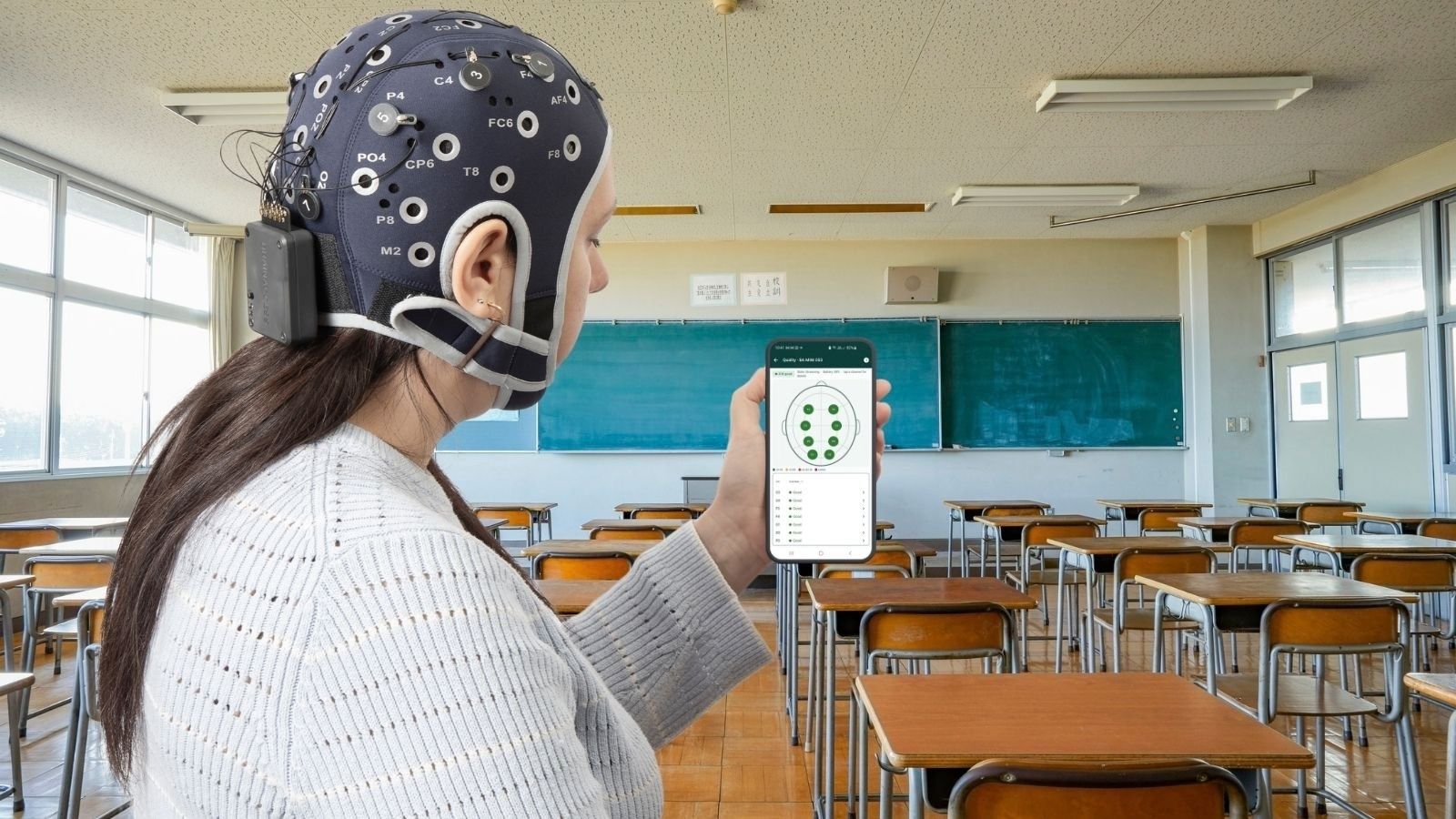
Fast forward to now, and EEG looks almost nothing like Berger’s setup:
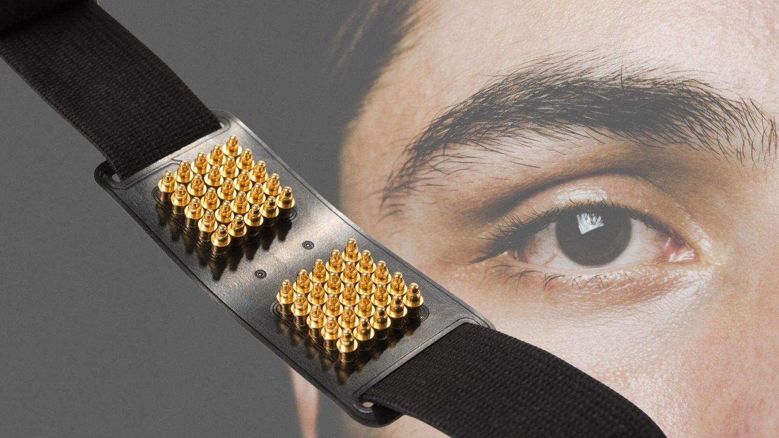
- Dry electrodes instead of gel.
- Wireless headsets instead of a wired galvanometer.
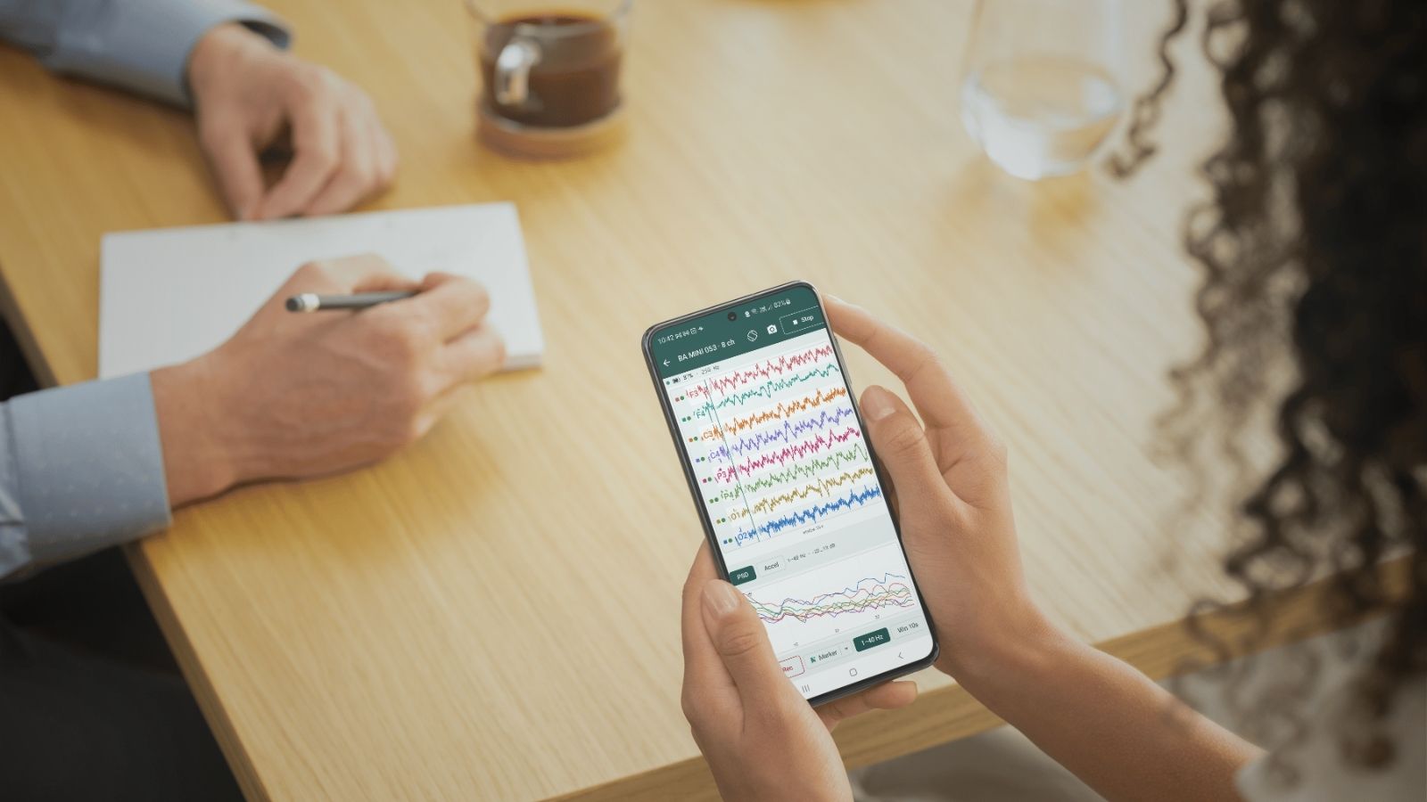
- Machine learning models trained on brain data instead of a researcher squinting at a paper trace.
- Labs run hyperscanning studies recording multiple brains simultaneously.
- Systems pair EEG with eye tracking, heart rate, and motion sensors.
None of it erases what Berger did in that room in Jena a century ago, it’s all still built on the same basic act: catching the brain’s electrical signal and trying to make sense of it.
This month, at BrainAccess, we are releasing Jena AI, our new AI assistant to help researchers actually make sense of their EEG setups.
Jena is built for the same reason Berger kept adjusting his equipment for five stubborn years: to make the signal a little easier to trust, and a little easier to use.
A hundred years after a flickering line on a strip of paper in Jena, the city’s name is on our software. Seemed only right.
Box 2: What is Jena AI?
Jena is an EEG-aware AI assistant built into the BrainAccess Board desktop app, currently free in beta. It keeps an eye on your connection and device status, checks data quality across multiple devices at once — handy if you’re running a hyperscanning or multimodal setup — and can walk you through Board’s settings and features when you’d rather ask a question than dig through a manual.
Reference
[1] Vergani, A. A. (2024). Hans Berger (1873–1941): the German psychiatrist who recorded the first electrical brain signal in humans 100 years ago. Advances in Physiology Education, 48(4), 878-881.
[2] Rossini, P. M., Cole, J., Paulus, W., Ziemann, U., & Chen, R. (2025). 1924–2024: First centennial of EEG. Clinical Neurophysiology, 170, 132-135.
[3] Haas, L. F. (2003). Hans berger (1873–1941), richard caton (1842–1926), and electroencephalography. Journal of Neurology, Neurosurgery & Psychiatry, 74(1), 9-9.
[4] “Berger Studies the Human Electroencephalogram.” EBSCO Research Starters. ebsco.com